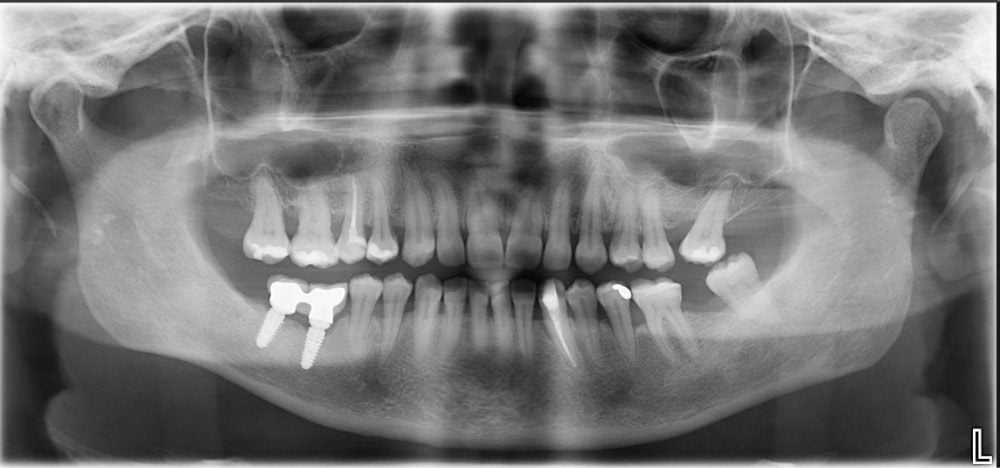
A precise diagnosis is the key to a successful treatment of dental illnesses, while X-ray imaging of the mouth is a fundamental part of it. In fact, only through an X-ray can dental practitioners have an insight into the state of the tooth, but also into that of the jaw bone and detect possible inflammation processes, all of which allows for a precise diagnosis and faster recovery. Nowadays, 2D and 3D images are regularly used as they visualise the tooth as well as the surrounding soft tissue, in fact the orthopantomogram or panoramic X-ray includes images of both jaws, the jaw articulation and the sinuses.
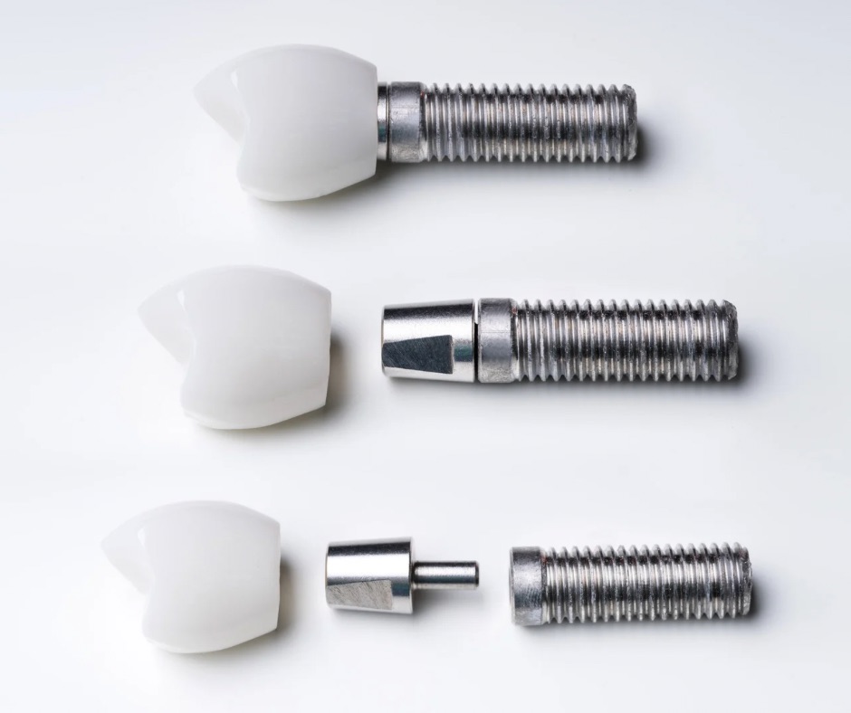
Thanks to digital methods it is possible to obtain a full image of the jaws in just a few minutes, with image quality far superior to that of the classic radiographic techniques. Moreover, this kind of imaging is particularly useful when planning surgery or implantological procedures.
How is the imaging done?
The device which is used to produce a digital panoramic X-ray rotates around the patient’s head emitting a beam of X rays. The procedure lasts around 30 seconds and the image is immediately available on a computer’s screen. The amount of radiation to which the patient is exposed is minimal, presenting no health hazard.
Riviera Dent offers digital panoramic radiography with a Planmeca device for both diagnosis and implantology planning purposes.